PMR has a classic pattern — aching and stiffness in both shoulders (often hips/thighs) with morning stiffness lasting longer than 45 minutes in adults 50 years old or older.
Because other problems can look similar, doctors rule out inflammatory arthritis, shoulder/hip disorders, thyroid and muscle diseases, drug effects (e.g., statins), infections, and cancer-related syndromes before confirming PMR. History + exam + ESR/CRP + targeted imaging (often ultrasound) sort this out.
Why the “mimics” matter
- Picking the wrong label can lead to the wrong treatment (e.g., unnecessary steroids in non-PMR conditions).
- Some mimics need urgent or different care (e.g., infection, malignancy, inflammatory myopathy).
- A structured approach avoids missed diagnoses and helps set expectations for recovery.
How clinicians approach the differential
- Start with the story: Age 50 or older, bilateral shoulder (± hip) aching, morning stiffness lasting over 45 minutes, function hits (coat sleeves, getting up from a chair).
- Inflammation labs: ESR/CRP usually high in PMR (but can be normal in a minority); use labs with the whole picture.
- Focused exam: Shoulders/hips (ROM limits from stiffness), check hands/wrists/knees (if inflamed, think elderly-onset RA), screen for red flags.
- Imaging when needed: Ultrasound of shoulders/hips can show bilateral bursitis/tenosynovitis supporting PMR; it also helps spot rotator-cuff problems.
- Avoid the “steroid trap”: a quick steroid response alone doesn’t prove PMR — other conditions can briefly improve on steroids.
Note: Doctors may refer to the 2012 EULAR/ACR classification criteria (and versions with ultrasound) to support the pattern, especially to distinguish PMR from RA and other inflammatory diseases. These criteria support (they don’t replace) clinical judgment.
A quick triage: symptoms that point away from PMR
- One-sided pain only;
- Numbness/tingling (nerve problem);
- True muscle weakness (not just stiffness);
- Hot, swollen small joints (hands/wrists) early on;
- Very high CK (muscle enzyme) or clear thyroid problem.
These cues push doctors to look for another cause first.
Top categories of PMR mimics (with “how to tell” tips)
1) Inflammatory arthritides
Elderly-onset rheumatoid arthritis (EORA) / seronegative RA
- Clues for EORA: swollen/tender hands, wrists, knees; may be seronegative (RF/anti-CCP negative) early; shoulder/hip aching overlaps with PMR.
- Tests/Imaging: RF/anti-CCP (may be negative), ultrasound or MRI of hands/wrists; erosions over time.
- Key difference: Peripheral joint inflammation is more typical; PMR’s hallmark is shoulder/hip girdle stiffness without prominent small-joint synovitis.
Spondyloarthritis (late-onset)
- Clues: inflammatory back pain, enthesitis (tendon insertions), psoriasis, IBD, uveitis.
- Imaging: sacroiliitis on MRI.
RS3PE (Remitting Seronegative Symmetrical Synovitis with Pitting Edema)
- Clues: sudden hand/foot swelling with pitting edema, seronegative.
- Course: steroid-responsive but a different entity; sometimes overlaps with malignancy or other rheumatic diseases.
CPPD (“pseudogout”) with shoulder/hip-girdle pattern
- Clues: flares, chondrocalcinosis on X-ray/ultrasound; can mimic PMR stiffness.
- Test: joint aspiration (if peripheral joint involved) shows CPPD crystals.
2) Shoulder and hip disorders (mechanical/degenerative)
Rotator cuff tears/tendinopathy; adhesive capsulitis (frozen shoulder)
- Clues: often one-sided, mechanical pain, night pain from a torn cuff; restricted passive ROM in frozen shoulder.
- Imaging: shoulder ultrasound or MRI shows cuff tear or capsular thickening.
- Why this matters: Ultrasound that shows bilateral SASD bursitis/tenosynovitis supports PMR, whereas a focal cuff tear points away.
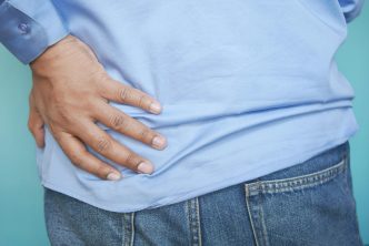
Hip osteoarthritis / lumbar spinal stenosis or radiculopathy
- Clues: groin pain (hip OA), pain with weight-bearing; leg numbness/tingling or walking-distance-limited calf pain (spine/stenosis).
- Imaging: X-ray/MRI as guided by exam.
3) Muscle and endocrine disorders
Inflammatory myopathies (polymyositis, dermatomyositis, inclusion-body myositis)
- Clues: true weakness (e.g., can’t rise from a chair even when pain controlled), high CK, sometimes rash (DM).
- Testing: CK, aldolase, myositis antibodies, EMG/MRI; biopsy if needed.
Hypothyroidism
- Clues: fatigue, weight gain, cold intolerance; normal CK or mildly high; aches and stiffness can mimic PMR.
- Test: TSH (± free T4).
Statin-associated muscle symptoms / drug-induced myopathy
- Clues: started or increased statin; proximal aches ± high CK; pattern may be more myopathy than stiffness.
- Action: never stop a statin on your own — discuss with your clinician; alternative causes must be checked.
4) Systemic illness and infection
Viral illnesses (parvovirus B19, EBV, hepatitis, HIV, etc.)
- Clues: recent viral-like illness, fever; usually self-limited; labs vary.
- Tests: targeted serologies as indicated by history.
Bacterial infection (including occult infection or endocarditis)
- Clues: fevers, sweats, weight loss, heart murmur, anemia; high inflammatory markers but atypical pain pattern.
- Tests: blood cultures, echo, imaging guided by symptoms.
Malignancy and paraneoplastic syndromes
- Clues: unexplained weight loss, night sweats, anemia, very high inflammatory markers without a fitting PMR pattern.
- Note: large series suggest a temporary bump in cancer detection near the time of PMR evaluation—likely from more testing and because some cancers mimic PMR at onset. Careful assessment at diagnosis is standard.
5) Neurologic and other
Parkinson disease and neurologic stiffness
- Clues: bradykinesia, rigidity, tremor; stiffness may be misread as musculoskeletal.
- Exam: neurologic signs guide referral.
Depression, obstructive sleep apnea, chronic pain syndromes
- Clues: global fatigue, non-restorative sleep, widespread pain disproportionate to exam; labs normal.
- Plan: screen and treat these conditions—they can mimic or magnify aches in older adults.
Not a mimic, but critical: Giant cell arteritis (GCA) is PMR’s “sister” disease. New headache, scalp tenderness, jaw pain with chewing, or any vision change is same-day urgent—doctors often start treatment before all tests return to protect vision.
Side-by-side quick guide
| Condition | Clues that favor it (vs PMR) | Helpful tests |
|---|---|---|
| Elderly-onset RA | Swollen/tender hands/wrists; morning stiffness but peripheral joints involved | RF/anti-CCP; hand/wrist US/MRI; erosions over time |
| Rotator-cuff disease | Often one-sided shoulder pain; pain with overhead reach; focal weakness | Shoulder US/MRI focal tear; PMR US tends to be bilateral bursitis |
| Hip OA / Lumbar stenosis | Groin pain (hip), weight-bearing pain; leg numbness/tingling or distance-limited calf pain (spine) | X-ray/MRI as indicated |
| Inflammatory myopathy | True weakness (proximal) more than stiffness; high CK; sometimes rash | CK/aldolase; myositis panel; EMG/MRI; biopsy |
| Hypothyroidism | Cold intolerance, weight gain, constipation; labs otherwise bland | TSH (± free T4) |
| Statin myopathy | New/worse aches after statin start or dose change; possible high CK | CK; med review; don’t stop without guidance |
| CPPD (“pseudogout”) | Flares; chondrocalcinosis | X-ray/US; crystal analysis if effusion present |
| RS3PE | Pitting edema of hands/feet; seronegative | Clinical pattern; screen for associations |
| Viral/infectious | Fever, recent illness, systemic signs | Targeted serologies, cultures/echo if endocarditis suspected |
| Fibromyalgia | Widespread pain, poor sleep, fatigue; normal ESR/CRP | 2016 ACR WPI/SS scoring; exclude other causes (context) |
Tests doctors often order (and why)
- ESR/CRP: inflammation markers — often high in PMR, very helpful to track; not perfectly specific.
- CBC, CMP: anemia or infection clues; kidney/liver baseline before meds.
- CK: screens for muscle damage (myositis, statin effects).
- TSH: rules out hypothyroidism.
- RF/anti-CCP: if RA is in the mix.
- Shoulder/hip ultrasound: supports PMR when it shows bilateral bursitis/tenosynovitis and helps rule out rotator-cuff tears.
Practical pitfalls (and how clinicians avoid them)
- Calling any shoulder pain “PMR.” Mechanical shoulder disease is common; ultrasound helps separate PMR from cuff tears/frozen shoulder.
- Relying on steroid response alone. Many conditions feel better briefly on steroids; we still need the pattern and appropriate testing.
- Missing RA or CPPD. Look for small-joint signs and consider crystal disease in older adults.
- Overlooking infection or malignancy. Unexplained fevers, weight loss, anemia, or very high markers push the work-up toward systemic causes.
When to escalate or seek a second opinion
- Atypical features (one-sided pain, true weakness, numbness/tingling, marked small-joint swelling).
- Normal ESR/CRP with a poor response to adequate steroid dosing.
- Relapses very early in tapering or side effects piling up.
Rheumatology review, targeted imaging, or broader labs are appropriate next steps.
Bottom line
PMR is one pattern among many causes of shoulder-hip girdle pain and morning stiffness in older adults. A careful combination of history, exam, labs (ESR/CRP, CK, TSH), and targeted imaging (ultrasound first) separates PMR from mimics such as EORA/seronegative RA, rotator-cuff disease, hip OA/spine disease, inflammatory myopathy, thyroid problems, statin myopathy, CPPD, infections, malignancy, and others. Getting this step right sets up the right treatment and a smoother recovery.
SOURCES
- 2012 EULAR/ACR PMR classification criteria—pattern and the role of ultrasound; helps distinguish from RA and other inflammatories. ard.bmj.com
- Cleveland Clinic Journal of Medicine updated review (2020): broad list of PMR mimics (EORA, spondyloarthritis, RS3PE, CPPD, others). CCJM
- bpacnz primary-care guide (2023): practical “look before you leap” checklist and work-up steps. Best Practice Advocacy Centre
- StatPearls (2024): differential includes hypothyroidism, viral infections, OSA, depression, and more. NCBI
- Medscape Differential (2023): adds endocarditis, dermatomyositis, malignancy, Parkinson disease, RS3PE. Medscape
- BMJ review (2008): highlights statin-related myalgia, neurologic causes, malignancy in the differential. PubMed