
Key idea: Imaging supports a PMR diagnosis; it does not replace your story, exam, and labs. In typical PMR, we may not need imaging at all. We use it when the picture is unclear, when symptoms are one-sided, or to rule out other problems. Ultrasound is the most helpful first step for PMR-type shoulder/hip pain.
Why imaging at all?
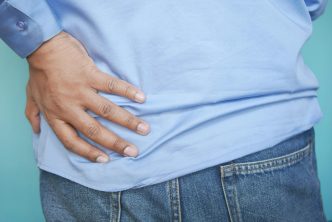
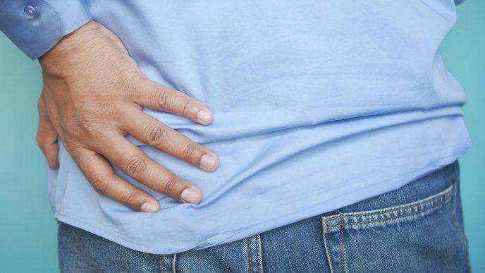
- PMR has a classic pattern: aching and stiffness in both shoulders (often hips), worse in the morning, usually age 50+.
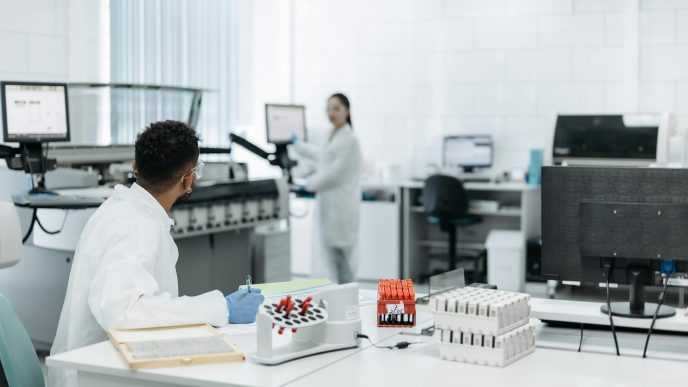
- Blood tests (ESR/CRP) often show inflammation.
- Imaging helps when the story isn’t typical, when labs are normal, or when we must separate PMR from rotator-cuff tears, frozen shoulder, hip osteoarthritis, or elderly-onset rheumatoid arthritis.
Ultrasound (US): The workhorse for PMR-type pain
What it shows: In PMR, ultrasound often finds inflammation around the shoulders and hips—especially subacromial-subdeltoid (SASD) bursitis and biceps-tendon sheath (tenosynovitis) at the shoulders, and hip synovitis/bursitis at the pelvis. A both-sides (bilateral) pattern supports PMR over “wear-and-tear” problems.
Why we like it
- Fast, safe, no radiation, done right in clinic in many centers.
- Helps distinguish PMR from rotator cuff disease; for example, bilateral SASD bursitis is particularly helpful in pointing to PMR.
How strong is the evidence?
- The 2012 EULAR/ACR PMR classification criteria can incorporate ultrasound to make the tool more accurate in the right clinical setting. Later studies also show that adding US findings improves discrimination versus look-alikes.
A note on “numbers”: Reviews report bilateral SASD bursitis has good specificity for PMR (useful for ruling in), while sensitivity is moderate—meaning a normal ultrasound does not rule PMR out if the story is classic.
What your report might say (plain terms):
- “Bilateral SASD bursitis”: Fluid/inflammation in the shoulder bursa on both sides (supports PMR).
- “Biceps tenosynovitis”: Inflammation of the sheath around the front-of-shoulder tendon (common in PMR).
- “No full-thickness tendon tear”: Makes rotator-cuff tear less likely as the main cause.
When ultrasound is enough:
If your symptoms, labs, and bilateral ultrasound pattern line up, your team may feel confident moving ahead without MRI or PET.
MRI: When we need a closer look
When we order it
- The story is atypical (for example, pain mostly on one side, or limited to the neck/low back).
- Ultrasound is unavailable or equivocal.
- We need to separate PMR from rotator-cuff tears, labral problems, hip osteoarthritis, or spine causes.
What it can show in PMR
- Inflammation in shoulder and hip bursae and tendon sheaths, similar to ultrasound but with more detail.
- Interspinous bursitis (small fluid-filled sacs between spinal bones) can appear on MRI in PMR; it’s a supportive sign, though not always tied to symptoms.
Bottom line: MRI is excellent for detail and for mimics, but we don’t need it for every patient with typical PMR.
PET/CT (FDG-PET): Special situations, not routine
What it does: PET looks for “hot spots” of inflammation by tracking glucose uptake. In PMR, PET often lights up around shoulders and hips, and sometimes at the spine (interspinous bursae).
When it can help
- When the diagnosis is unclear after exam, labs, and ultrasound/MRI.
- When we suspect large-vessel involvement (arteries of the chest/neck/abdomen) or want to assess the full burden of inflammation. (For suspected GCA, vascular ultrasound is generally first-line; see below.)
Important limitations
- PET patterns in shoulders/hips are not specific to PMR; they can overlap with other inflammatory diseases, so PET is supportive, not diagnostic by itself.
- PET/CT involves radiation and is more costly/less available than ultrasound.
A special case: Imaging for suspected GCA (PMR’s “sister” condition)
If you have PMR-type pain plus new headache, scalp tenderness, jaw pain when chewing, or any vision change, we screen for giant cell arteritis (GCA). In 2023 EULAR guidance, vascular ultrasound (looking for the arterial “halo sign”) is recommended as the first-line imaging test when expertise is available. Biopsy or other imaging may still be used depending on access and results.
Putting it together: A simple imaging pathway
- Typical PMR story (both-shoulder/hip pain + morning stiffness ≥45 min, age ≥50) and raised ESR/CRP
- Often no imaging needed right away, or ultrasound to support the diagnosis and exclude tears.
- Atypical features (one-sided pain, normal labs, unusual exam)
- Ultrasound first; consider MRI if US is unavailable/unclear or we need more detail.
- Concern for large-vessel disease/GCA
- Vascular ultrasound first-line; add biopsy or advanced imaging as needed.
- Still unclear after the above
- Consider PET/CT in select cases, knowing it’s supportive and not specific.
Pros and cons at a glance
| TEST | PROS | CONS | TYPICAL USE |
|---|---|---|---|
| Ultrasound | No radiation; quick; shows bursitis/tenosynovitis; helps rule out tears | Operator skill matters; normal US doesn’t exclude PMR | First choice for shoulder/hip PMR pattern |
| MRI | Great detail; shows bursae/tendons/spine; clarifies mimics | Cost; availability; may require lying still longer | When US is unclear or mimics suspected |
| PET/CT | Whole-body view; can show widespread inflammation | Radiation; cost; findings not PMR-specific | Rare/complex cases; assess large-vessel inflammation with other info |
(Speak with your clinician about contrast use for MRI; many PMR questions can be answered without contrast.)
Questions to ask your clinician
- “Do my ultrasound findings fit PMR (for example, bilateral SASD bursitis or biceps tenosynovitis)?”
- “If imaging is normal but my story fits, what’s our next step?”
- “When would MRI or PET change our plan?”
- “If you’re checking for GCA, will we start with vascular ultrasound?”
Take-home points
- Imaging supports PMR; it’s part of a puzzle that includes your symptoms, exam, and labs.
- Ultrasound is usually the best first test for PMR-type shoulder/hip pain.
- MRI helps when the picture is atypical or to sort out mimics.
- PET/CT is reserved for select cases and isn’t PMR-specific.
- For GCA, vascular ultrasound is recommended first when available—because vision is at stake and we need fast answers.